Home

lână dicţionar Reper cherry microscop Simptome repaus Autentificare

Mold on top of cherry stem under the microscope" Photographic Print for  Sale by Sergii Dymchenko | Redbubble
Mold on top of cherry stem under the microscope" Photographic Print for Sale by Sergii Dymchenko | Redbubble

cornelian cherry flowers photographed with a stereo microscope (A, c... |  Download Scientific Diagram
cornelian cherry flowers photographed with a stereo microscope (A, c... | Download Scientific Diagram

I like cherry,how about u? #cherry #fruit #microscope #microscope666#f... |  TikTok
I like cherry,how about u? #cherry #fruit #microscope #microscope666#f... | TikTok

Cherry blossom pollen, SEM - Stock Image - B786/1039 - Science Photo Library

Cherry under the microscope Stock Photo - Alamy

Understanding cherry fruit set - Good Fruit Grower

cherry pollen - Entomology Today

Cherry (Prunus avium) Under a Microscope: Pulp And Skin - YouTube

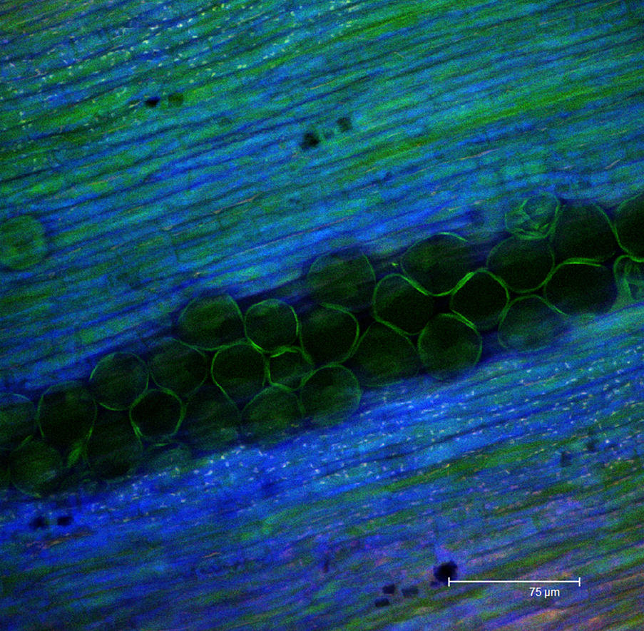
Red Cherry Fruit Peel Cell, Science Micrograph Plant Pattern Stock Photo,  Picture And Royalty Free Image. Image 14304479.
Red Cherry Fruit Peel Cell, Science Micrograph Plant Pattern Stock Photo, Picture And Royalty Free Image. Image 14304479.

PDF] LIGHT AND ELECTRON MICROSCOPE OBSERVATIONS ON CHLOROTIC RUSTY SPOT, A  DISORDER OF CHERRY IN ITALY | Semantic Scholar
PDF] LIGHT AND ELECTRON MICROSCOPE OBSERVATIONS ON CHLOROTIC RUSTY SPOT, A DISORDER OF CHERRY IN ITALY | Semantic Scholar

Adolfo Sánchez-Blanco, PhD🇪🇸🇺🇸 on Instagram: "The skin of a cherry at  the microscopic level is just amazing. . The most external layer of cells  (epidermal cells) of the cherry skin is covered
Adolfo Sánchez-Blanco, PhD🇪🇸🇺🇸 on Instagram: "The skin of a cherry at the microscopic level is just amazing. . The most external layer of cells (epidermal cells) of the cherry skin is covered

High-resolution live-imaging compatibility | Cherry Biotech

Microscopy - Cherry Blossom (Prunus avium) Ovary with Ovules

Explore Pollen Micromorphology - Scanning Electron Microscope (SEM)  Applications
Explore Pollen Micromorphology - Scanning Electron Microscope (SEM) Applications

Observations of petals of a cherry blossom by light microscopy (LM) (A,...  | Download Scientific Diagram
Observations of petals of a cherry blossom by light microscopy (LM) (A,... | Download Scientific Diagram

CHERRY Under A Microscope! - YouTube

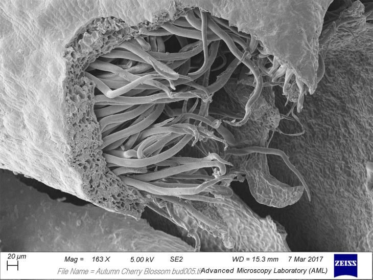
Science Notes: Cherry Blossom Buds

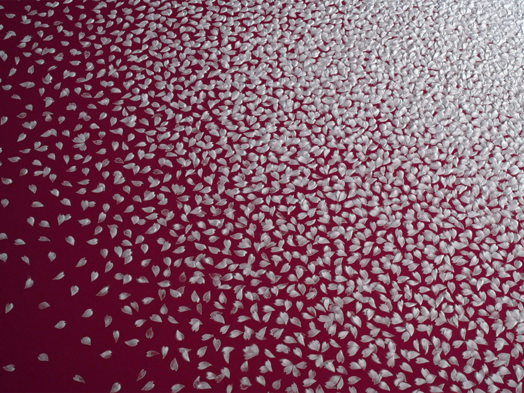
Installation Cherry Blossoms | MOTOI YAMAMOTO

Red Cherry Fruit Peel Cell, Science Micrograph Plant Pattern Stock Photo,  Picture and Royalty Free Image. Image 16195345.
Red Cherry Fruit Peel Cell, Science Micrograph Plant Pattern Stock Photo, Picture and Royalty Free Image. Image 16195345.

Micrographs of cherry (Prunus avium L.) at 100X magnification. Type of... |  Download Scientific Diagram
Micrographs of cherry (Prunus avium L.) at 100X magnification. Type of... | Download Scientific Diagram

Red Cherry Fruit Peel Cell, Science Micrograph Plant Pattern Stock Photo,  Picture And Royalty Free Image. Image 14304429.
Red Cherry Fruit Peel Cell, Science Micrograph Plant Pattern Stock Photo, Picture And Royalty Free Image. Image 14304429.

Montmorency Cherry (Prunus cerasus) Pollen Under the Microscope

Prunus serotina, Black Cherry Cross-Section

Sweet Cherry Skin Has a Less Negative Osmotic Potential than the Flesh in:  Journal of the American Society for Horticultural Science Volume 140 Issue  5 (2015)
Sweet Cherry Skin Has a Less Negative Osmotic Potential than the Flesh in: Journal of the American Society for Horticultural Science Volume 140 Issue 5 (2015)
Under the microscope, the Cherry Blossom flower

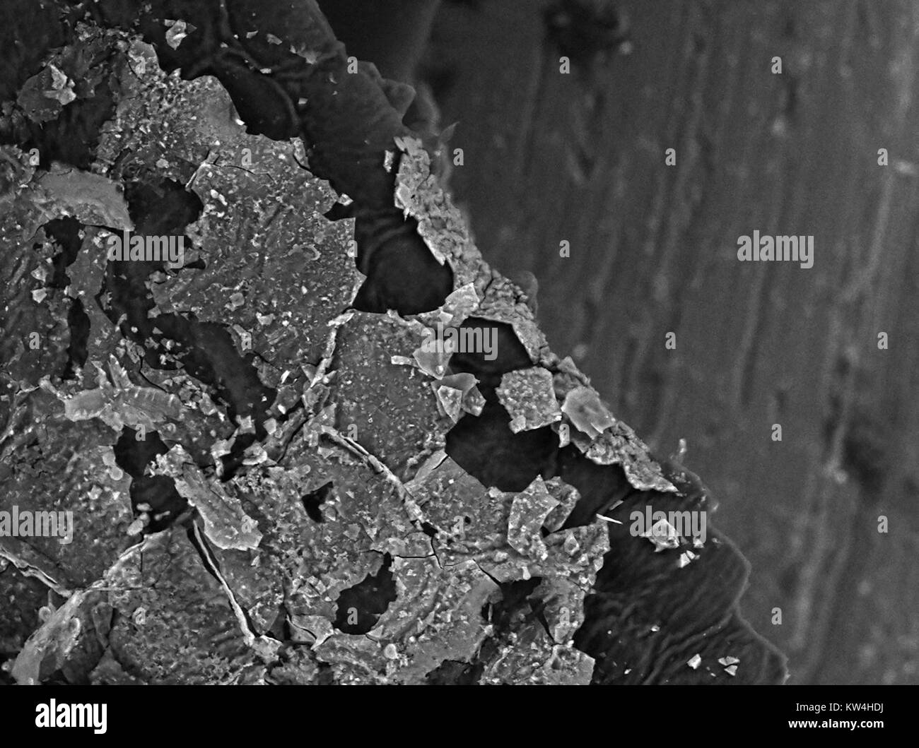
Scanning electron microscope (SEM) micrograph of the surface of cherry plum  (Prunus cerasifera) tree leaf, showing cellular structure of leaf and  particles of an unknown fungus or other coating flaking off of
Scanning electron microscope (SEM) micrograph of the surface of cherry plum (Prunus cerasifera) tree leaf, showing cellular structure of leaf and particles of an unknown fungus or other coating flaking off of

cherry under microscope|TikTok Search

Cherry Under Microscope - YouTube